Feeding and Respiration
Elysia sp. is a member of the sacoglossa, a specialist group of opisthobranchs that feed in a unique suctorial manner that is often considered to be borderline parasitic
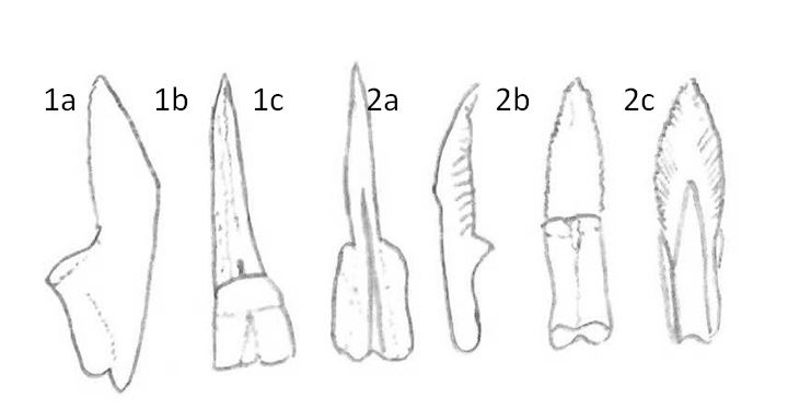
[2,26]. Their specialist teeth pierce the cell walls of their algal prey and suck out the cytoplasm of the cell in a highly efficient feeding strategy [27]. The radula is composed of two aspects, an ascending aspect where the new teeth form, and a descending aspect that leads into the ascus sac [1]. As a result of their unusual feeding strategy their radula teeth wear out quite quickly, and in fact the sacoglossa, formerly ascoglossa, derive their name from the ascus sac into which they deposit used teeth [27]. Figure 1 below illustrates the type of uniserial radula seen in the sacoglossans.
It is thought that the sacoglossa evolved in close association with the filamentous algae Caulerpa and have now diversified to feed on a variety of food types including fleshier algae such as Halimeda, diatoms and even the eggs of other animals [27]. Many species are known to be specialists feeding on only one algal species [2], though Elysia sp. appeared to feed on both Chlorodesmis sp. and Halimeda sp. over the course of this study. The video below shows one of the animals collected for this study browsing over the rubble, presumably in search of food.
Video- Elysia sp. browsing coral rubble in search of food.
Unusually, two functional 'photosynthetic' systems are seen within the opisthobranchia. The sacoglossans are able to incorporate and maintain functional chloroplasts in their digestive systems, while the nudibranchs are known to form symbioses with zooxanthellae, similar to those seen in the corals [1]. The Elysiids' incorporation of functional chloroplasts acquired from their algal prey into their own tissues is quite unique amongst the animal kingdom [1,2,15,27-31]. Known as kleptoplasty, this process occurs in two forms throughout the sacoglossa, with non-functional kleptoplasty commonplace, and functional kleptoplasty only seen within the Elysiidae [27,28]. Non-functional kleptoplasty is usually considered to be the uptake and short-term retention of chloroplasts within the digestive tract, mostly for camouflage purposes [27,28]. Functional kleptoplasty is generally defined as the uptake of chloroplasts that are then used for photosynthetic purposes within the slug, and are retained for a longer duration, in some cases up to several months [27,28]. In order to increase the efficiency of this energy acquiring strategy, the Elysiidae have co-opted the generous surface area afforded by their parapodia into a respiratory organ.
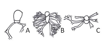
As they respire across their entire dorsal surface, the shell-less sacoglossans, including the Elysiidae, have extensively branched dorsal vessels that tend to be quite prominent, albeit highly variable between groups [1,2]. These lead to the pericardial prominence located posterior to the head, and can be used as a diagnostic characteristic to distinguish between groups, and even between species as is indicated in Figure 2 below [1,2,27]. The entire epithelial surface is also covered in microvilli to increase nutrient uptake in the sacoglossans [2]. As a consequence of the mechanism by which these slugs respire, feed and acquire energy, they are limited in their distribution both globally and on a local scale to shallow water environments where light attenuation, oxygen concentration and nutrient levels are high. As such, they are also particularly susceptible to extinction as a result of habitat alteration, please see the Conservation and Threats page for further information on the threats posed to the sacoglossa.

Figure 1- Schematic diagrams of the uniserial radula of the Elysiidae. Diagrams 1a-c are of a blade-shaped tooth drawn from a lateral (a), frontal (b) and posterior (c) view . Diagrams 2a-c are of a tricuspid tooth with lateral projections drawn from a lateral (a), frontal (b) and posterior (c) view. Diagram adapted from Jensen, 1992 [1].

Figure 2- The extent to which the dorsal vessels cover the upper surface of the parapodia varies significantly between species. A, Elysia timida; B, Elysia trisinuata; C, Elysia ornata. Diagrams adapted from Jensen, 1992 [1].
|